Services
All About Joint Fusion
Arthritis of the foot and ankle is a common and often debilitating condition in which the protective cartilage covering the joint surfaces becomes worn or damaged, leading to pain, stiffness, swelling, and progressive loss of function. When conservative measures — including footwear modification, orthotic insoles, physiotherapy, and injection therapy — no longer provide adequate relief, surgery may offer a lasting solution.
Fusion Surgery — known medically as arthrodesis — is one of the most reliable and well-established surgical treatments for end-stage arthritis in the foot. By permanently joining the surfaces of an arthritic joint, fusion eliminates the painful movement between worn surfaces, relieves pain, and restores stability. Whilst the joint no longer moves independently after fusion, the foot can compensate well through the surrounding joints, and the majority of patients achieve a significant improvement in their quality of life.
Conditions Treated with Fusion Surgery
Big Toe Arthritis
First metatarsophalangeal joint fusion (hallux rigidus)
Hallux rigidus is arthritis of the joint at the base of the big toe and is the most common form of arthritis in the foot. It causes progressive stiffness and pain at the big toe joint, particularly with walking, climbing stairs, or pushing off during activity. In its earlier stages it may respond well to conservative measures and injection therapy, but in advanced disease with significant joint destruction, fusion offers the most reliable and durable outcome.
First metatarsophalangeal joint (MTPJ) fusion involves removing the remaining damaged cartilage and holding the joint in an optimal functional position — slightly elevated and turned outwards — using a combination of screws and sometimes a plate. Once fused, the toe is stable and pain free, and the remaining joints of the foot adapt well to the loss of movement at this level.
First MTPJ fusion is considered the gold standard surgical treatment for advanced hallux rigidus and is associated with high rates of patient satisfaction and return to comfortable walking. Most patients are able to walk in a post-operative shoe from day one and return to normal footwear by eight to twelve weeks.
Midfoot & Hindfoot Arthritis
The midfoot comprises a collection of small joints that together provide the arch and stability of the foot. Arthritis in this region — whether arising from wear and tear, previous injury, inflammatory arthritis, or flatfoot deformity — can cause a deep, aching pain across the top or inner aspect of the midfoot that is often worse with prolonged standing or walking on uneven ground.
The Naviculocuneiform Joints
Where the navicular bone meets the three cuneiform bones — and the talonavicular joint — between the talus and the navicular at the apex of the arch — are among the most important midfoot articulations and are commonly affected by arthritis. Because of their role in arch mechanics, arthritis here can also contribute to progressive flatfoot deformity.
Fusion of one or more of these joints stabilises the midfoot, corrects any deformity, and relieves pain reliably in well-selected patients. The joints are prepared and held in their corrected positions using screws or staples whilst the bones heal together, typically over a period of ten to fourteen weeks. Because the midfoot joints contribute relatively little independent movement in normal gait, their fusion is generally well tolerated functionally.
Pre-operative diagnostic injection into the affected joint or joints is an important step in the assessment of midfoot arthritis. Because several joints lie in close proximity, a targeted injection with local anaesthetic helps to confirm precisely which joint is the primary pain source before surgical fusion is planned.
Subtalar Arthritis
Subtalar joint fusion
The subtalar joint sits beneath the ankle joint proper, between the talus and the calcaneus (heel bone). It is responsible for the side-to-side rocking motion of the hindfoot — the movement that allows the foot to adapt to uneven terrain. Subtalar arthritis causes deep pain on the outer and inner aspects of the hindfoot, often described as a grinding or aching sensation that worsens with walking on slopes, steps, or uneven ground, and with twisting movements of the foot.
It most commonly arises following trauma — particularly calcaneal fractures — but can also occur as a result of inflammatory arthritis, adult-acquired flatfoot deformity, or generalised wear. It is sometimes mistaken for ankle joint arthritis, and careful examination and imaging are required to identify it as the primary source of symptoms.
Subtalar fusion involves preparing the joint surfaces and compressing the talus and calcaneus together, holding them in a position of neutral alignment with screws whilst the bones fuse. Where there is associated hindfoot deformity — such as valgus or varus malalignment — this is corrected at the time of surgery. The ankle joint above is preserved and continues to function normally, and most patients find that the loss of subtalar movement is well compensated by the rest of the hindfoot and foot complex.
A pre-operative diagnostic injection into the subtalar joint under fluoroscopic or ultrasound guidance is a valuable step in confirming the diagnosis before surgery is planned, particularly where ankle joint arthritis or other hindfoot pathology may also be present.
What Fusion Surgery Involves
Pre-Operative Planning
Weight-bearing X-rays and CT or MRI scanning are used to assess the extent of joint damage, identify any associated deformity, and plan the surgical approach and fixation. Diagnostic injection may be performed in advance to confirm the affected joint. Your general health, bone quality, and any factors affecting healing — such as smoking, diabetes, or steroid use — are also carefully assessed, as these can influence fusion rates.
The Procedure
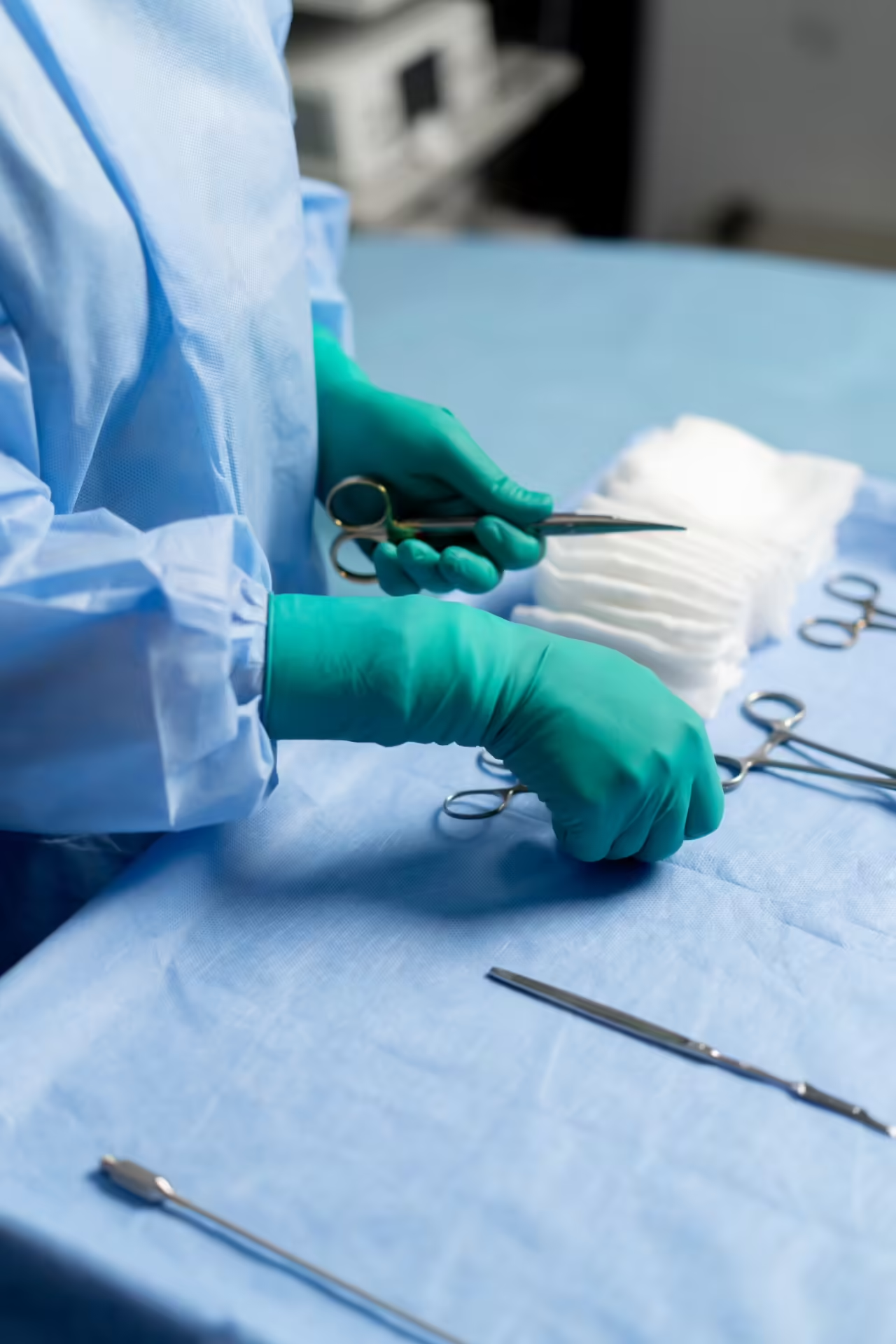
Fusion surgery is carried out under regional or general anaesthetic. The joint is accessed, the remaining cartilage and damaged bone surfaces are carefully prepared, and the bones are positioned and compressed together in their optimal alignment. Rigid internal fixation — using screws, plates, or staples depending on the joint — holds everything securely in place whilst the bones heal and knit together. The procedure is performed as a day case or with a one-night stay depending on the complexity.
Recovery & Rehabilitation
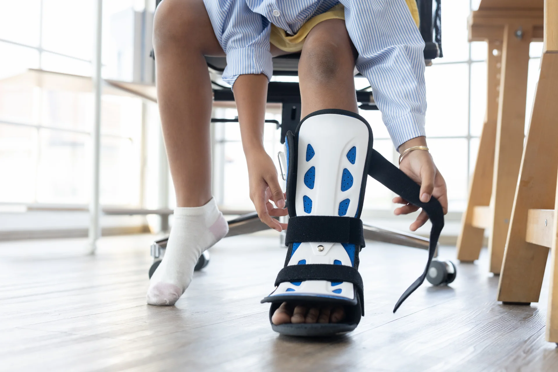
Recovery following fusion surgery requires a period of protected weight-bearing whilst the bones heal together. Depending on the joint fused and the surgical technique used, this typically involves a period in a plaster cast or boot, followed by progressive return to weight-bearing and normal footwear. Physiotherapy is an essential component of recovery, helping to restore strength, balance, and walking pattern.
Big toe fusion
Walk in post-op shoe from day one. Normal shoes 8–12 weeks. Full activity 4–6 months.
Midfoot fusion
Non-weight-bearing 6–8 weeks. Boot to 12 weeks. Normal shoes 3–4 months.
Subtalar fusion
Non-weight-bearing 6–8 weeks. Boot to 12 weeks. Normal shoes 4–6 months.
Benefits, Risks & Complications
- Reliable, durable pain relief
- Correction of associated deformity
- Restoration of stable, comfortable walking
- High long-term patient satisfaction
- Avoids need for ongoing injections or medication
- Wound infection or breakdown
- Deep vein thrombosis (DVT)
- Nerve or vessel injury
- Swelling, bruising, stiffness
- Anaesthetic complications
- Non-union — failure of the bones to fuse
- Malunion — fusion in a suboptimal position
- Hardware irritation requiring screw removal
- Transfer arthritis in adjacent joints over time
- Incomplete pain relief
Non-union risk: Certain factors are known to increase the risk of non-union following fusion surgery. These include smoking, poorly controlled diabetes, osteoporosis, and obesity. I will discuss these factors with you at consultation and advise on steps that can be taken to optimise your chances of a successful fusion.
Fusion is a permanent procedure and the decision to proceed should never be taken lightly. I will always ensure that conservative options have been thoroughly explored and that the diagnosis has been confirmed before recommending surgery. A clear and honest discussion of what you can realistically expect — including the recovery demands and the permanent nature of the procedure — is an essential part of the consultation process.