What We Treat
All About Bunions
A bunion — known medically as hallux valgus — is one of the most common foot conditions seen in podiatric surgical practice. Despite how familiar the term is, bunions are frequently misunderstood, and many patients spend years managing pain and discomfort without knowing that effective, long-lasting treatment is available. This page explains what a bunion is, why it develops, and the full range of treatment options I offer — from conservative measures through to surgery.
What is a Bunion?
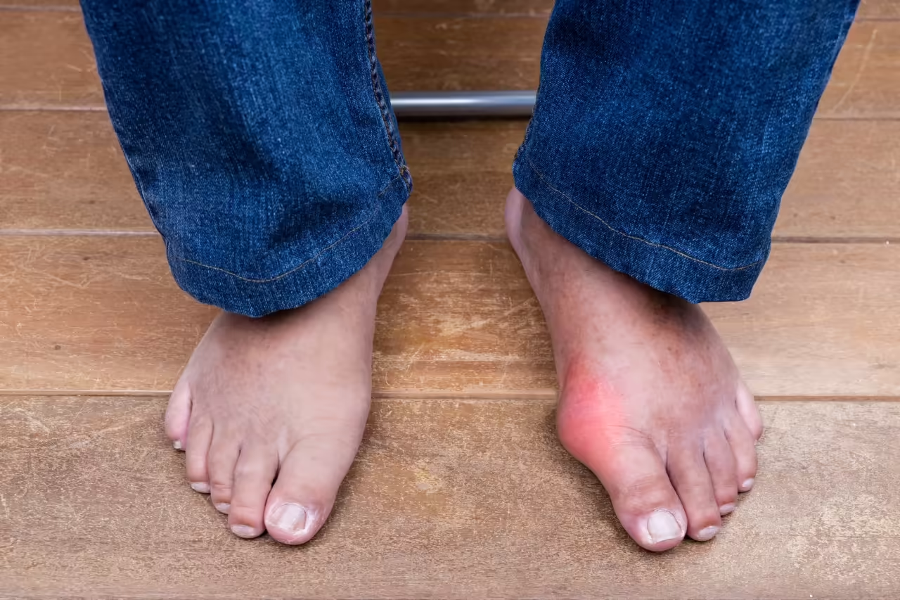
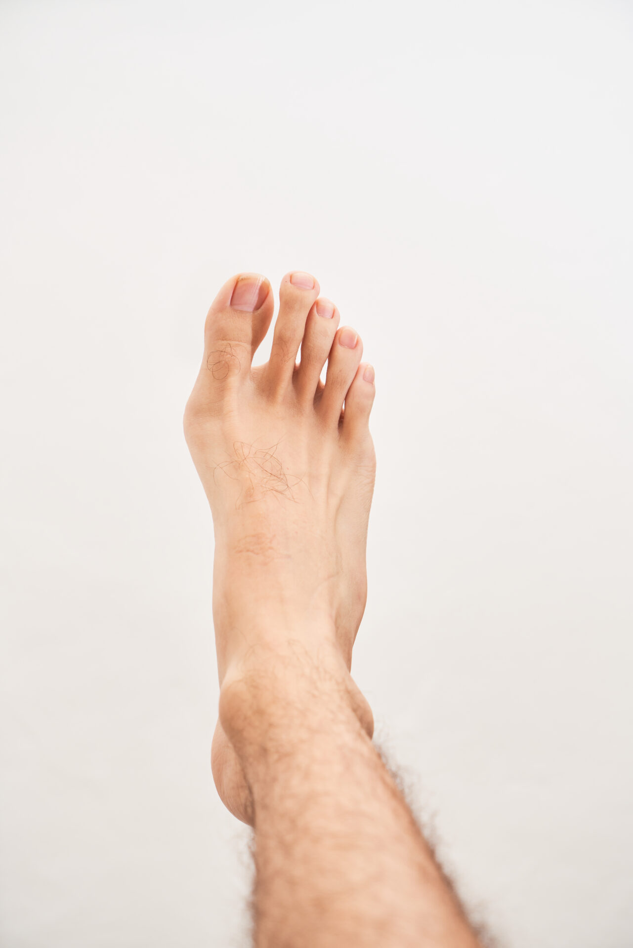
A bunion is a progressive deformity of the first metatarsophalangeal joint — the joint at the base of the big toe. The condition involves two related changes: the first metatarsal bone drifts inwards (towards the midline of the body) whilst the big toe drifts in the opposite direction, towards the lesser toes. This creates the characteristic bony prominence on the inner side of the foot at the base of the big toe, which is the bump most people recognise as a bunion.
Over time, the joint becomes increasingly misaligned. The soft tissues around the joint — tendons, ligaments, and the joint capsule — adapt to the abnormal position, which can make the deformity self-perpetuating. In more advanced cases the big toe may cross over or under the second toe, the lesser toes may develop secondary deformities, and the joint itself may become arthritic.
Bunions are significantly more common in women than men, though they do affect both sexes. They tend to worsen gradually over time and rarely improve without treatment.
What Causes Bunions?
Bunions are multifactorial — meaning several factors contribute to their development. The most important of these is heredity. Bunions run strongly in families, and the underlying bone architecture and foot mechanics that predispose to the deformity are largely inherited. If your mother or grandmother had bunions, you are at significantly higher risk of developing them yourself.
Hereditary Factors
The shape of the foot, the mobility of the first metatarsal, and the ligamentous laxity that predispose to bunion formation are largely genetically determined.
Footwear
Narrow, pointed, or high-heeled shoes do not cause bunions but can accelerate progression and worsen symptoms in those who are already predisposed.
Foot Mechanics
Hypermobility of the first ray, flatfoot deformity, and abnormal pronation can all contribute to the instability and drift of the first metatarsal that drives bunion formation.
Inflammatory Arthritis
Conditions such as rheumatoid arthritis can accelerate joint destruction and deformity at the first MTPJ, leading to more rapid and severe bunion development.
Symptoms
Not all bunions are painful — some patients have a significant deformity with relatively mild symptoms, whilst others have a smaller bunion that causes considerable discomfort. Typical symptoms include pain and tenderness over the bony prominence, inflammation and redness of the overlying skin, difficulty finding comfortable footwear, restricted or painful movement of the big toe joint, and pressure pain on the ball of the foot from the altered mechanics of the forefoot. In more advanced cases, pain may be present even at rest.
It is worth noting that the degree of deformity does not always correlate with the degree of pain — treatment decisions are based on the impact your bunion is having on your life, not purely on the size of the bump.
Non-Surgical Management
Conservative measures will not correct a bunion or prevent it from worsening over time, but they can help to manage symptoms and slow progression. They are always the appropriate starting point for patients whose symptoms are mild or who are not yet ready for surgery.
Footwear Modification
Wide-fitting shoes with a broad toe box reduce pressure on the bunion and are the single most effective conservative measure.
Orthotic Insoles
Custom insoles can help to control abnormal foot mechanics and offload the first metatarsal head, reducing pain during activity.
Padding & Splinting
Protective padding over the bunion prominence reduces friction and irritation. Toe spacers and night splints may offer comfort, though they do not correct the deformity.
Surgical Treatment
Surgery for bunions has evolved considerably in recent years and today’s techniques offer reliable correction, faster recovery, and better cosmetic outcomes than ever before. I offer the full range of surgical options for hallux valgus, from modern minimally invasive keyhole procedures through to more complex reconstructive surgery for severe deformity and arthritis. The procedure I recommend will be based on a careful assessment of your X-rays, the severity of your deformity, the health of your joint, and your personal goals and lifestyle.
All bunion surgery is aimed at achieving the same fundamental goals: removing the bony prominence, realigning the first metatarsal and big toe, and restoring a balanced, pain-free foot. How this is achieved depends on the procedure selected.
Minimally Invasive Bunion Surgery (MIS)
Keyhole technique
Minimally invasive bunion surgery — often referred to as keyhole surgery — is a modern technique in which the bony correction is performed through incisions of just a few millimetres. Specialised small instruments and a fine burr are used under continuous X-ray guidance (fluoroscopy) to remove the prominence and perform a precise osteotomy — a controlled cut of the metatarsal bone — which allows it to be repositioned and held in place with small titanium screws.
MIS offers significant advantages over traditional open surgery: less soft tissue disruption, reduced post-operative swelling and pain, minimal scarring, and the ability to walk immediately in a post-operative shoe from day one in most cases. Recovery is generally faster than with open techniques, and patients typically return to normal footwear by six to eight weeks and to full activity within three to four months.
MIS is suitable for mild to moderate hallux valgus and selected cases of more severe deformity. It is performed as a day case under local or general anaesthetic and is the technique I offer most frequently for primary bunion correction in suitable patients.
Open Bunion Surgery
Traditional open bunion surgery remains an excellent and well-proven technique, and is the preferred approach for certain deformity patterns, revision cases, or where the anatomy is not suited to a minimally invasive approach. Through a carefully placed incision along the inner side of the foot, the metatarsal is cut and repositioned, the soft tissues are rebalanced, and the prominent bone is removed. The corrected position is held with screws or a plate.
A wide range of osteotomy techniques exist for open bunion correction — each suited to different deformity severities and bone geometries. The choice of technique will be discussed with you in detail at consultation based on your X-ray findings. Recovery following open surgery sometimes involves a slightly longer period in a post-operative shoe compared to MIS, though functional outcomes are excellent and long-term satisfaction rates are high.
For Severe or Hypermobile Deformity
In patients with a severe bunion, a significantly widened forefoot, or underlying hypermobility of the first ray — where the first metatarsal is excessively mobile at its base — a standard osteotomy alone may not provide sufficient or durable correction. In these cases, the Lapidus procedure offers a more definitive solution.
The Lapidus procedure involves fusing the first tarsometatarsal joint — the joint at the very base of the first metatarsal where it meets the medial cuneiform bone — in a corrected position. By stabilising the root cause of the instability rather than simply correcting the metatarsal shaft, the Lapidus addresses the underlying hypermobility that drives recurrence in susceptible patients. It is considered the most powerful correction available for hallux valgus and is associated with very low recurrence rates.
The joint is held securely in its corrected position using a low-profile plate and screws whilst the fusion consolidates, typically over eight to twelve weeks. Modern Lapidus techniques — including minimally invasive variants — allow earlier weight-bearing than some traditional approaches, and outcomes in appropriately selected patients are excellent.
Recovery following a Lapidus procedure is somewhat longer than after a standard osteotomy, reflecting the time needed for the fusion to consolidate. A period of protected weight-bearing is required, and return to normal footwear is typically achieved by three to four months, with full return to activity by six months.
First Metatarsophalangeal Joint Fusion
For severe deformity or arthritis
For patients with a severe bunion deformity complicated by significant osteoarthritis of the first metatarsophalangeal joint, first MTPJ fusion — also known as arthrodesis — is the most appropriate and reliable surgical option. This procedure permanently joins the bones of the big toe joint in an optimally functional position, eliminating both the deformity and the painful arthritic joint surface in a single operation.
First MTPJ fusion is also the procedure of choice for patients with a very severe bunion where the joint has become irreversibly unstable or where previous bunion surgery has failed and further osteotomy is not feasible. It is a well-established procedure with a strong evidence base and consistently high patient satisfaction, particularly in active individuals who place significant demands on their feet.
The joint is held in its corrected, fused position using a combination of screws and a dorsal plate. Once fused, the big toe is stable and pain free. Whilst the joint no longer moves, the overall function of the foot is well preserved — most patients are able to walk, exercise, and wear a wide range of footwear including many styles of normal shoe.
Most patients can walk in a post-operative shoe from day one. Return to normal footwear is typically achieved by eight to twelve weeks, and full return to activity by four to six months.

How Do I know Which Procedure Is Right For Me?
The decision about which surgical technique is most appropriate for you is made on an individual basis, taking into account the severity of the deformity as measured on weight-bearing X-rays, the presence or absence of joint arthritis, the mobility and stability of the first ray, your age, activity level, bone quality, and your personal goals for treatment.
At your consultation I will review your imaging, examine your foot in detail, and explain clearly which procedure I recommend and why. I will also discuss realistic expectations for the outcome and recovery, so that you can make a fully informed decision in your own time without any pressure.