What We Treat
All About Flatfoot
Flatfoot is one of the most common and complex conditions affecting the foot and ankle. It encompasses a spectrum of problems ranging from a mildly low arch that causes no symptoms at all, through to a severely collapsed and painful foot that significantly affects mobility and quality of life. Understanding what is causing your flatfoot — and how far along the spectrum your condition sits — is essential to planning the right treatment.
I have a specialist interest in the assessment and surgical management of flatfoot deformity and offer the full range of procedures — from minimally invasive implant surgery through to complex reconstructive osteotomy and fusion — tailored to the individual patient and the stage of their condition.
What is Flatfoot — And What Is It Called?
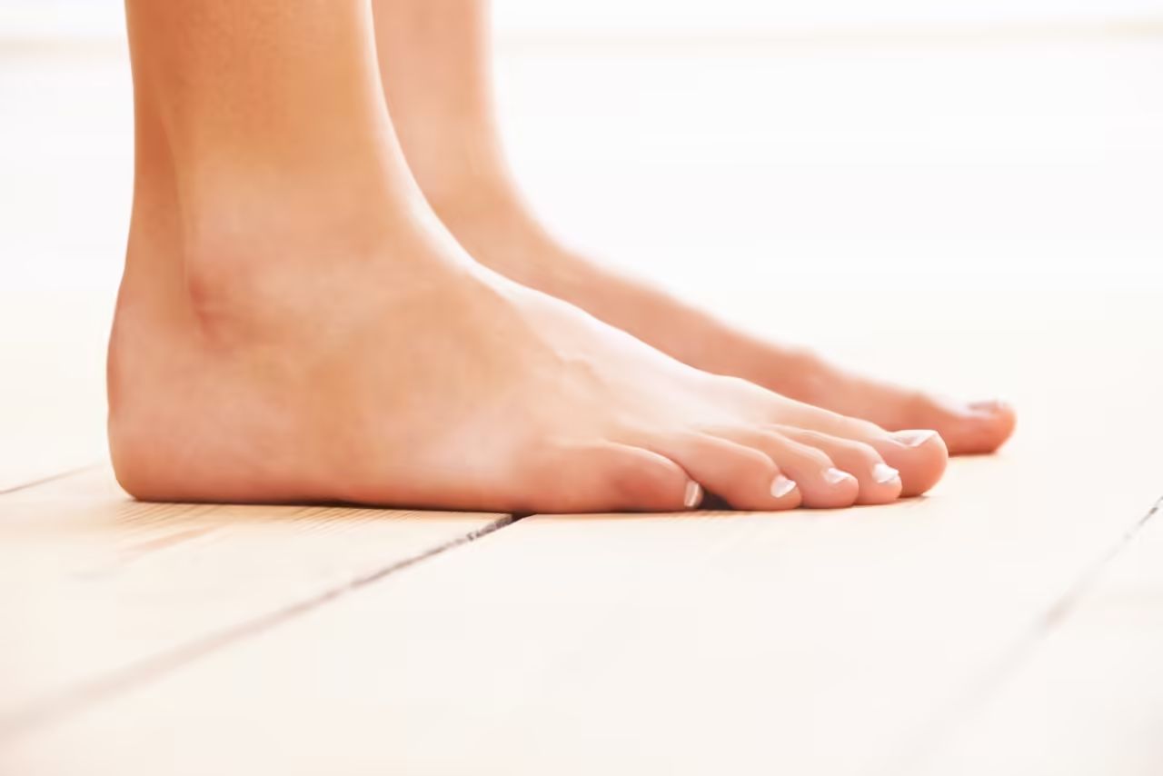
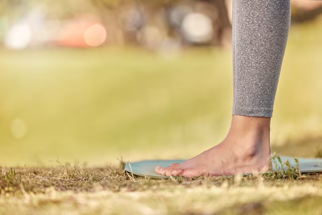
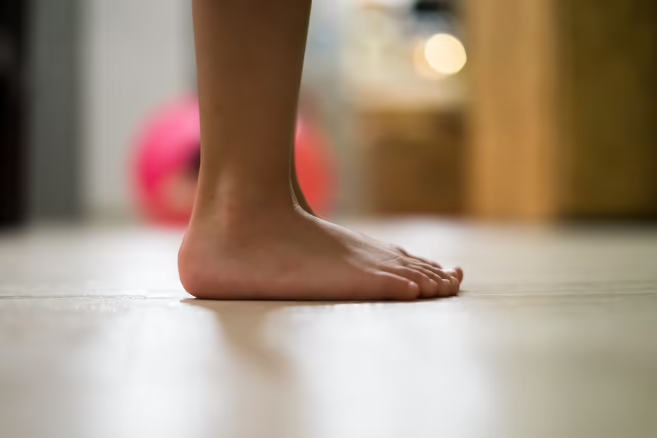
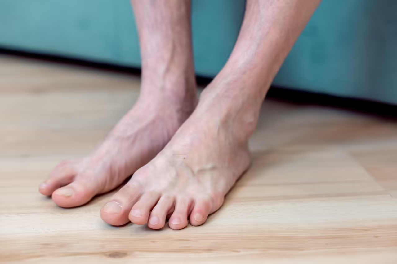
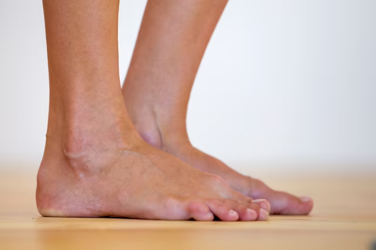
Flatfoot is a condition in which the arch of the foot is reduced or absent, causing the inner border of the foot to collapse towards or contact the ground. The heel typically rolls outward (hindfoot valgus), the midfoot sags, and in more advanced cases the forefoot splays and turns outward relative to the hindfoot.
The condition goes by several names, which can be confusing for patients. You may have heard it referred to as:
Pes Planus
The Latin medical term meaning “flat foot,” used to describe the characteristic low or absent arch.
Adult-Acquired Flatfoot
A term emphasising that the deformity has developed in adulthood rather than being present from birth, most commonly due to tibialis posterior tendon dysfunction.
Progressive Collapsing Foot Deformity (PCFD)
The most recently adopted terminology, reflecting that the condition involves multiple structures collapsing across the whole foot — not just the arch — and that it is progressive if untreated.
Tibialis Posterior Tendon Dysfunction (TPTD)
A term referring to the most common underlying cause in adults — failure of the tibialis posterior tendon, the primary dynamic supporter of the arch.
Throughout this page, I will use the term flatfoot for simplicity, though I may use these other terms when discussing specific aspects of the condition.
Symptoms
Symptoms vary considerably depending on the stage and severity of the deformity. In early stages, patients may notice aching along the inner ankle and arch, particularly after prolonged standing or walking, and a gradual change in the shape of the foot. As the deformity progresses, pain becomes more constant, the ankle may feel weak or unstable, and shoes wear out asymmetrically. In advanced stages, pain extends to the outer ankle as the collapsing foot impinges on the lateral structures, and significant functional limitation is common.
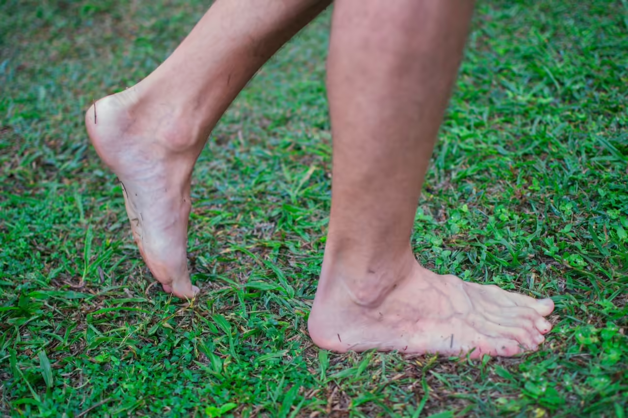
A characteristic sign is that the affected foot cannot perform a single-leg heel rise — standing on the toes on one foot — as the weakened tibialis posterior tendon is unable to lock the hindfoot for this movement.
Non-Surgical Management
Conservative management is always the first line of treatment, particularly in earlier stages of the condition. Whilst it cannot correct a structural deformity once established, it can control symptoms effectively and in some cases slow progression. For patients who are not surgical candidates, conservative management is the mainstay of long-term care.
Orthotic Insoles
Custom or semi-custom arch-supporting insoles are the cornerstone of conservative management. They support the medial arch, control hindfoot valgus, and reduce the load on the tibialis posterior tendon.
Ankle-Foot Orthosis (AFO)
For more advanced deformity or acute tibialis posterior tendon dysfunction, a rigid or semi-rigid AFO provides greater control of the hindfoot and ankle, reducing pain and limiting further collapse.
Physiotherapy
Strengthening of the tibialis posterior, intrinsic foot muscles, and calf complex, combined with calf stretching to address equinus, forms the basis of a structured rehabilitation programme.
Injection Therapy
Targeted corticosteroid injection into the tibialis posterior tendon sheath or subtalar joint can reduce acute inflammation and pain. Injection directly into the tendon substance is avoided due to the risk of rupture.
Important: Flatfoot deformity is progressive. Conservative measures can manage symptoms but cannot reverse established structural collapse. Early surgical intervention, when appropriate, generally produces better outcomes than operating at a very advanced stage when deformity is severe and joints have become arthritic.
Surgical Treatment
Surgery for flatfoot is tailored precisely to the stage and pattern of each individual’s deformity. Because a flatfoot deformity involves multiple structures — the tendon, the ligaments, the bones, and the joints — surgical correction often involves a combination of procedures performed together to address every component of the collapse. The procedures I offer are described below.
Surgery is performed under regional or general anaesthetic. Most procedures require a period of non-weight-bearing followed by progressive rehabilitation, and a commitment to the full recovery programme is essential to achieving the best outcome.
Calcaneal Osteotomy
A calcaneal osteotomy involves making a precise surgical cut through the heel bone and shifting it into a corrected position to realign the hindfoot. In flatfoot reconstruction, the most commonly performed technique is a medialising calcaneal osteotomy, in which the back of the heel bone is shifted inward to correct the characteristic outward rolling (valgus) of the heel. This directly offloads the tibialis posterior tendon and restores more normal alignment through the hindfoot and ankle.
The repositioned bone is held securely with a screw or plate whilst it heals. A calcaneal osteotomy is one of the most powerful tools available for hindfoot correction and forms the cornerstone of many flatfoot reconstructions, typically performed in combination with other soft tissue or bony procedures.
Achilles Tendon & Gastrocnemius Lengthening
Tightness of the calf muscle complex — known as equinus — is present in the majority of patients with a flatfoot deformity and is a major driver of arch collapse. A tight calf forces the foot into abnormal pronation with every step, placing excessive strain on the arch-supporting structures and accelerating the deformity.
Addressing equinus is therefore an essential component of flatfoot surgery. I perform either a gastrocnemius recession — release of the gastrocnemius muscle at the level of the calf — or a formal Achilles tendon lengthening, depending on the level and severity of the tightness. Gastrocnemius recession is generally preferred where tightness is isolated to this muscle, as it preserves overall calf strength better than formal tendon lengthening.
Failing to address equinus at the time of flatfoot reconstruction is a well-recognised cause of recurrence and poor outcomes, and its assessment and treatment forms a standard part of my surgical planning.
Lateral Column Lengthening
In severe flatfoot deformity, the forefoot often abducts — swings outward relative to the hindfoot — producing the characteristic “too many toes” sign when viewed from behind. This forefoot abduction cannot always be corrected by hindfoot realignment alone and requires lengthening of the lateral column of the foot.
This is achieved through a calcaneal neck osteotomy or calcaneocuboid joint fusion — a cut through the front of the heel bone into which a bone graft or implant is inserted, pushing the outer column of the foot forward and correcting the outward swing of the forefoot. This procedure effectively reconstitutes the arch from the outside and is one of the most powerful arch-restoring procedures available.
It is typically used in combination with a other procedures to achieve a balanced, comprehensive reconstruction.
Medial Column Fusion
In cases where the arch collapse is driven primarily by instability or arthritis of the medial column joints — particularly the naviculocuneiform or talonavicular joints — fusion of one or more of these joints stabilises the inner arch and corrects the sagging midfoot.
Medial column fusion is particularly indicated where the midfoot has become arthritic as a consequence of the deformity, or where ligamentous disruption is so severe that bony stabilisation is needed to maintain arch correction achieved by other procedures. The joints are prepared and fused in a corrected, elevated position, restoring the arch height and providing a stable foundation for the foot.
In more advanced cases involving the talonavicular joint, fusion here provides powerful correction of both the hindfoot and midfoot deformity simultaneously, though it comes with a longer recovery and greater functional implications than more peripheral fusion procedures.
Plantar flexion osteotomy of the medial cuneiform
When the medial column of the foot is dorsiflexed — meaning the inner forefoot is elevated relative to the hindfoot — the arch lacks height even after hindfoot correction has been achieved. A plantar flexion osteotomy of the medial cuneiform (Cotton osteotomy) addresses this by opening a wedge in the medial cuneiform bone and inserting a graft or implant, forcing the inner forefoot downward and restoring the normal tripod contact pattern of the foot.
This procedure completes the medial arch reconstruction where the forefoot is not sitting correctly even after the hindfoot has been realigned, and is an important component of comprehensive flatfoot correction in patients where forefoot dorsiflexion is a significant part of the deformity.
Subtalar Implant (Arthroereisis)
Subtalar arthroereisis involves the insertion of a small implant into the sinus tarsi — a naturally occurring space on the outer side of the hindfoot between the talus and the calcaneus. The implant acts as a mechanical block, limiting the inward collapse of the talus and preventing excessive pronation of the hindfoot without fusing or permanently altering any joint.
The procedure is minimally invasive, reversible, and associated with a much shorter recovery than reconstructive osteotomy surgery. It is best suited to patients with flexible flatfoot — where the arch can still be reconstituted passively — and in younger patients or those who are not yet ready for or suitable for more extensive reconstruction. It is frequently used in combination with soft tissue procedures such as gastrocnemius recession and tibialis posterior tendon repair to provide a more complete correction.
If the implant causes persistent pain or discomfort in the sinus tarsi — a recognised complication — it can be removed relatively straightforwardly. This reversibility is one of the key advantages of arthroereisis over osteotomy-based correction.
Benefits, Risks & Recovery
Benefits
- Correction of the underlying structural deformity
- Significant and durable pain relief
- Prevention of further progression and joint arthritis
- Improved balance, stability, and walking function
- Reduced dependence on orthotics and bracing
General Surgical Risks
- Wound infection or breakdown
- Deep vein thrombosis (DVT)
- Nerve or vessel injury
- Anaesthetic complications
- Swelling, stiffness, and bruising
Procedure-Specific Risks
- Non-union or delayed healing of osteotomies
- Sinus tarsi pain from subtalar implant
- Hardware irritation requiring removal
- Incomplete correction or recurrence
- Adjacent joint arthritis over time
- Overcorrection producing a high-arched foot
Recovery Overview
- Arthroereisis: walking within days, normal shoes 6–8 weeks
- Osteotomy procedures: non-weight-bearing 6–8 weeks, boot to 12 weeks
- Normal shoes typically from 3–4 months
- Full recovery and return to sport: 9–12 months for complex reconstruction
- Physiotherapy throughout recovery is essential
A commitment to recovery: Flatfoot reconstruction is amongst the most involved surgery I perform, and recovery demands a significant commitment from the patient. A thorough discussion of what the surgery involves, what the recovery requires of you, and what outcome you can realistically expect is an essential part of the consultation process. I will never recommend surgery unless I am confident that the benefits clearly outweigh the risks for your individual situation.
If you have been told you have flat feet and are experiencing pain, instability, or difficulty with activity, I would encourage you to seek a specialist assessment. Many patients are surprised to learn that effective surgical options exist that can genuinely restore the shape and function of their foot. Early assessment, before the deformity becomes severe or the joints become arthritic, generally leads to a less complex procedure and a better outcome.